Immuno-(Dot)-Blot for the detection of the paraneoplastic autoantibodies anti-HuD, anti-Yo, anti-Ri, anti-CV2 (CRMP5),
anti-Amphiphysin, anti-Ma1 and anti-Ma2
Generally, these autoantibodies have been identified by immunohistochemical techniques. Due to problems with specificity a positive result in immunohistochemistry needs to be confirmed by an immunoblot, employing crude extracts from neuronal tissue as antigen. For example the reactivity of anti-Ro-antibodies, associated with Sjögren's-Syndrome, with protein bands resembling anti-HuD-immunoreactivity in crude extracts from neuronal tissue has been described. Immunohistochemistry is also laborious and requires a high degree of experience for reliable interpretation.
Figure 1: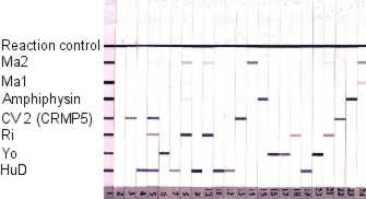
1 positive control
2 negative serum sample
3-24 different positive serum samples
Sensitivity and Specificity
47 positive serum samples, which were confirmed by IFT and other tests, were tested. Among these were 22 HuD-, 6 Yo-, 8 Ri-, 4 CV2-, 3 Amphiphysin- and 4 Ma2-positiv serum samples. All these samples were clearly identified as positive. To test spezificity, 46 serum samples of healthy persons, as well as patients showing other neurological symptoms, were tested. These samples showed no reaction with the antigens.
| Table: | Paraneoplastic neurological syndromes | Most frequently associated tumors |
| Anti-Hu-Antibodies(ANNA-1) |
|
Small-cell-lung cancer Non-small-cell lung cancer Extrapulmonary small cell cancer |
| Anti-Yo-Antibodies(Purkinje-cell-antigen) |
|
Breast cancer Ovarian cancer Uterus cancer |
| Anti-Ri-Antibodies(ANNA-2, anti-Nova-1) |
|
Breast cancer Small-cell-lung cancer Medullary carcinoma of the thyroid gland |
| Anti-CV2-(CRMP5-) Antibodies |
|
Small-cell-lung cancer Thymom |
| Anti-Amphiphysin-Antibodies |
|
Breast cancer Small-cell-lung cancer |
| Anti-Ma1 and Anti-Ma2- (Ta-) Antibodies |
|
Testicular cancer Lung-cancers |
* Brainstem encephalitis and cerebellar ataxia usually associated with tumors other than testicular and immunoreactivity against Ma2 and Ma1 proteins
Short description of test performance:
- Serum samples are diluted 1:2,000 in ready to use sample dilution buffer.
- Incubate the strips with 2 ml of the diluted serum specimen for 60 minutes at room temperature on a rocking table.
- wash five times with diluted wash buffer.
- Add 2 ml alkaline phosphatase IgG conjugate, ready to use, per strip.
- Incubate for 30 minutes at room temperature on a rocking table.
- wash five times with diluted wash buffer.
- Incubate each strip in 2 ml ready to use substrate-solution.
- Incubate for 25 minutes at room temperature until the bands become clearly visible. See control scan for comparison.
- Transfer the strips to distilled water to stop the reaction. Put the strips onto filter paper and let them dry. Store the strips in the dark.
back